
Brain MRI reveals no small blood vessel damage in hATTR, study finds
Routine scan offers no relevant diagnostic information in hATTR
Written by |
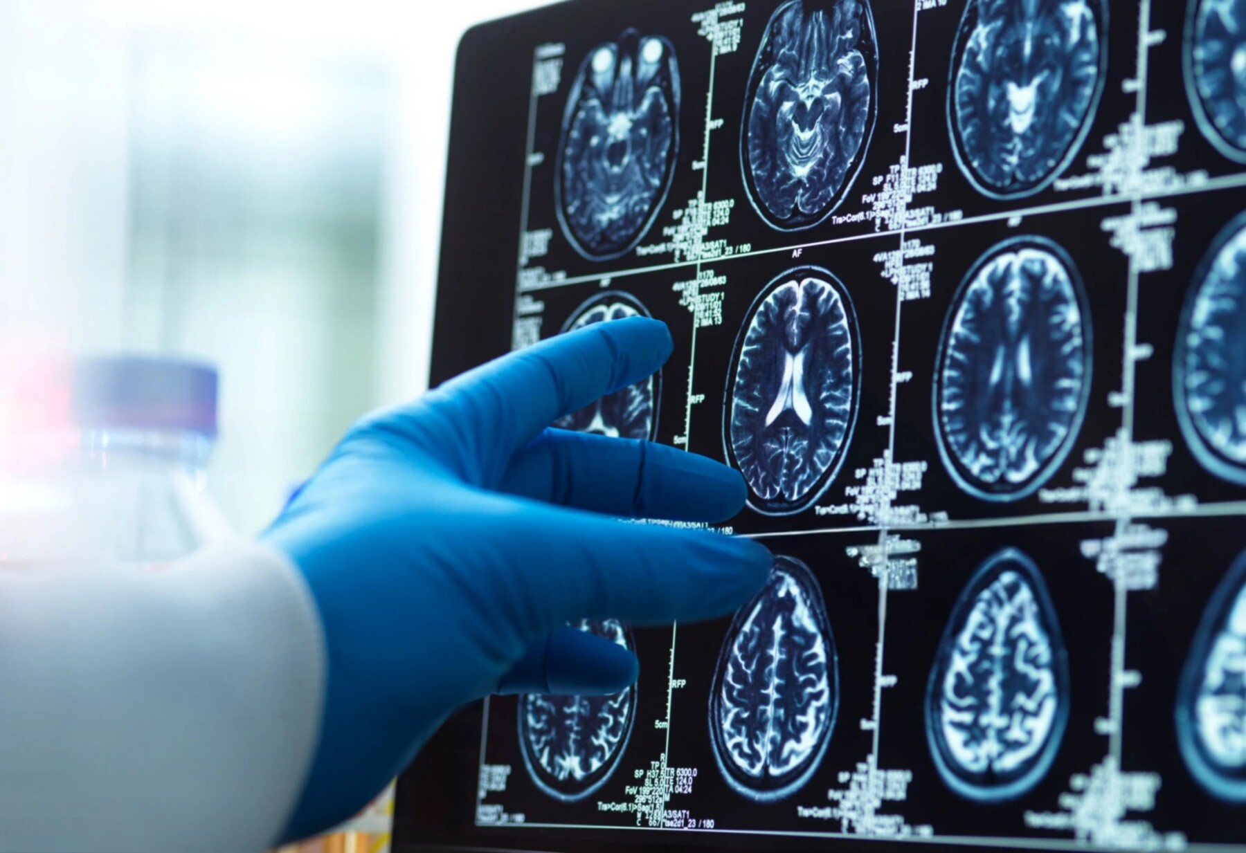
Hereditary transthyretin amyloidosis does not cause damage to small blood vessels in the brain that is visible on MRI scans, according to a study.
Hereditary transthyretin amyloidosis (hATTR), a group of conditions including hATTR with polyneuropathy (hATTR-PN) and hereditary ATTR cardiomyopathy (hATTR-CM), does not cause damage to small blood vessels in the brain that is visible on MRI scans, according to a study.
The single-center study in Italy involved 25 hATTR patients and seven people carrying disease-causing mutations but not yet showing symptoms (asymptomatic carriers).
“Taken together, these observations indicate that routine brain MRI does not provide relevant diagnostic information in the standard clinical evaluation of [hATTR] patients and should not be viewed as a useful diagnostic tool or red-flag indicator for disease activity,” researchers wrote.
The study, “No evidence of cerebral microangiopathy in hereditary transthyretin amyloidosis: An MRI case-control study from an Italian Center,” was published in the Journal of the Neurological Sciences.

‘Brain MRI appears to have little utility in routine diagnostic workup’
hATTR, also known as ATTRv, is caused by mutations in the gene that provides instructions to make a protein called transthyretin. The mutated protein forms toxic clumps, called amyloid deposits, that damage tissues and organs over time.
When amyloid deposits accumulate mainly in the peripheral nerves, or those outside the brain and spinal cord, the disease is referred to as hATTR-PN. When they build up in the heart, it is called hATTR-CM. Some patients have a mixed form of the disease, with damage to both the peripheral nerves and the heart.
While the neurological involvement in hATTR has been traditionally identified as a peripheral nervous system disease, increasing evidence supports significant involvement of the central nervous system (CNS), or the brain and spinal cord.
“Neuroimaging assessment of CNS involvement using brain magnetic resonance imaging (MRI) has yielded preliminary evidence of [blood vessel] brain injury in hATTRv,” the researchers wrote. However, “no robust characterization of the prevalence, severity, or distribution of these lesions across the ATTRv population has yet been established.”
Currently available treatments can effectively prevent the formation of toxic transthyretin clumps, but they can’t get into the brain. As such, brain involvement in hATTR-PN “is likely to become an increasingly important clinical concern for long-term survivors and may represent a critical frontier for future disease-modifying strategies,” the researchers wrote.
Brain MRI appears to have little utility in routine diagnostic workup or early disease screening.
In this study, a team of scientists in Italy evaluated whether people with hATTR show evidence of cerebral microangiopathy, or damage to the small blood vessels in the brain. To find out, they analyzed MRI data from 25 people with hATTR and seven asymptomatic carriers, as well as 32 age- and sex-matched people without the disease (used as controls).
Most hATTR patients (68%) had predominant heart involvement (hATTR-CM), while the remaining one-third had predominant involvement of peripheral nerves (hATTR-PN). The most common disease-causing mutation, present in five patients, was Ile88Leu.
Across various measures of cerebral microangiopathy, results showed that the hATTR group (both patients and asymptomatic carriers) showed no significant differences relative to the control group. There were also no significant differences between hATTR patients and controls and between people with hATTR-PN and those with hATTR-CM.
In addition, MRI-based measures showed no clear association with measures of disease severity in the overall hATTR group and in hATTR patients.
Altogether, even though prior studies have shown that toxic transthyretin clumps can form in the brain, this study suggests these clumps “do not translate into a clinically relevant or radiologically measurable small-vessel disease,” the researchers wrote. As such, “brain MRI appears to have little utility in routine diagnostic workup or early disease screening.”
Nonetheless, the researchers advocated for continual studies into how the brain may be impacted in hATTR, noting that there may be important changes that simply aren’t visible on MRI scans.




Leave a comment
Fill in the required fields to post. Your email address will not be published.